
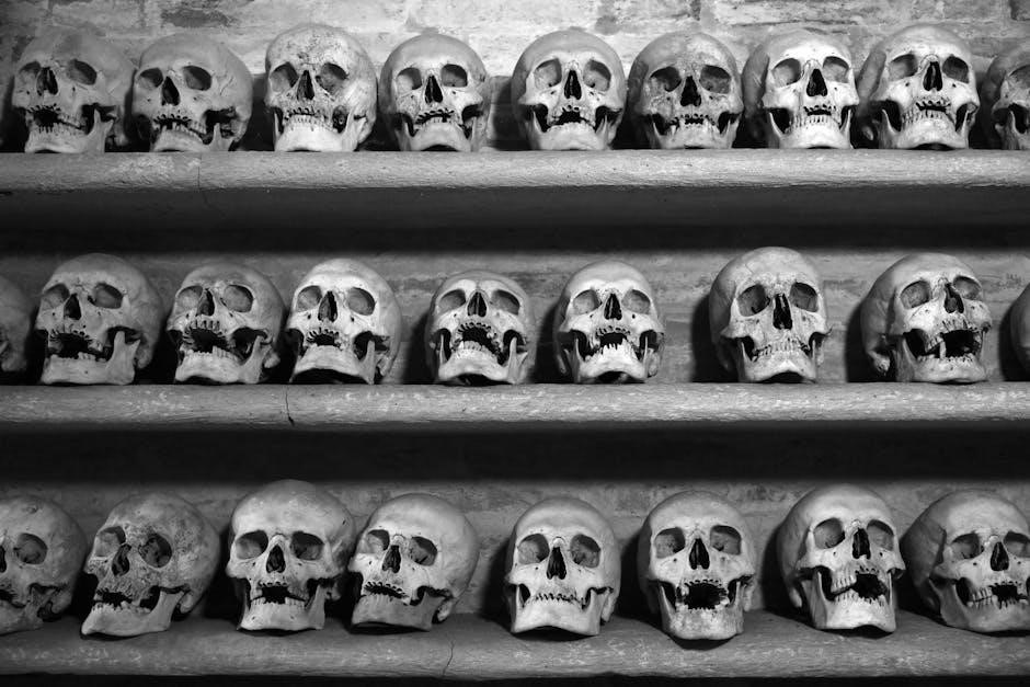
The femur, or thigh bone, is the longest and strongest bone in the human body. It connects the hip and knee, supporting body weight and enabling locomotion.
1.1 Overview of the Femur Bone
The femur, or thigh bone, is the longest, strongest, and heaviest bone in the human body, playing a critical role in the lower limb. It is a long bone, extending from the hip to the knee, and is classified as part of the appendicular skeleton. The femur is unique due to its robust structure, which enables it to support body weight and facilitate movement. Its proximal end forms a ball-and-socket joint with the pelvis, while the distal end articulates with the tibia and patella to form the knee joint. The femur is divided into three main parts: the proximal end (head, neck, and trochanters), the shaft (diaphysis), and the distal end (condyles and epicondyles). The head of the femur fits into the acetabulum of the pelvis, while the shaft provides attachment points for powerful muscles. The greater and lesser trochanters are prominent bony projections that serve as sites for muscle attachment, aiding in hip and knee movement. The femur’s unique anatomy makes it essential for locomotion and weight-bearing activities.
1.2 Location and Function of the Femur
The femur is located in the thigh, extending from the hip joint to the knee joint. It is the only bone in the thigh and serves as a critical structure for supporting the body’s weight and facilitating movement. The femur is positioned vertically in the lower limb, connecting the pelvis to the tibia and patella. Its proximal end is situated within the pelvic cavity, forming a ball-and-socket joint with the acetabulum, while its distal end interacts with the tibia and patella to form the knee joint. The femur’s primary function is to transmit forces from the trunk to the lower limbs, enabling activities such as walking, running, and standing. It also acts as a lever for the muscles of the hip and thigh, providing mechanical advantage for movement. The femur’s location and structure allow it to bear significant loads while maintaining flexibility and mobility, making it indispensable for human locomotion and stability.
1.3 Significance of the Femur in Human Anatomy
The femur holds profound significance in human anatomy due to its role as the longest, strongest, and heaviest bone, providing essential structural and functional support. It serves as the primary load-bearing bone, distributing body weight from the pelvis to the knees during activities like walking and running. The femur’s unique anatomy, including its robust shaft and specialized joints, allows it to withstand immense mechanical stress while maintaining flexibility. Additionally, the femur is a key attachment site for numerous muscles, tendons, and ligaments, enabling a wide range of movements such as hip flexion, extension, and rotation. Its importance extends beyond locomotion; the femur is also crucial for maintaining posture and balance. In forensic science, the femur’s length and structure are used to estimate stature and determine sex, making it a valuable tool in anthropological studies. Overall, the femur is a vital component of the human skeletal system, indispensable for both mobility and stability.

Structure of the Femur
The femur is a long bone comprising a head, neck, shaft, and distal end. Its strong shaft supports body weight, while the head forms a ball-and-socket joint with the hip, enabling mobility.
2.1 Proximal End of the Femur
The proximal end of the femur includes the head, neck, and trochanters. The head forms a ball-and-socket joint with the pelvis, while the neck connects the head to the shaft. The greater and lesser trochanters serve as attachment points for major hip muscles, facilitating movement.
2.2 Shaft of the Femur
The shaft of the femur is a long, cylindrical structure that connects the proximal and distal ends. It is robust and slightly curved, being convex anteriorly and concave posteriorly. The linea aspera, a prominent longitudinal ridge, reinforces the shaft and serves as an attachment site for several muscles and ligaments. Proximally, the shaft narrows and transitions into the neck, while distally, it widens, forming the supracondylar region. The shaft’s surface is smooth, with minimal muscle attachments directly along its length, allowing for efficient movement and weight distribution. Its strong cortical bone composition ensures durability, making it capable of withstanding significant stress and load. The shaft plays a critical role in maintaining posture and facilitating locomotion by acting as the primary structural support between the hip and knee joints.
2.3 Distal End of the Femur
The distal end of the femur is the thickest and most complex part of the bone, designed for weight distribution and joint stability. It consists of two rounded condyles (medial and lateral) that articulate with the tibia and patella, forming the knee joint. These condyles are covered in hyaline cartilage, facilitating smooth movement. Above the condyles lie the medial and lateral epicondyles, which serve as attachment points for ligaments and muscles, enhancing joint stability. The distal femur also features the supracondylar ridge, a bony prominence that strengthens the bone. Its wide, flattened structure allows for even weight distribution and supports various movements, such as walking and running. The distal end is crucial for absorbing and transmitting forces from the lower limb, making it essential for locomotion and overall mobility.

Anatomical Angles of the Femur
The femur has several key anatomical angles, including the femoral neck angle, femoral-tibial angle, and inclination angle, which play crucial roles in movement, weight distribution, and joint stability. These angles vary with age and gender, influencing gait and posture.
3.1 Femoral Neck Angle
The femoral neck angle, also known as the angle of inclination, is the angle between the femoral neck and shaft. It averages approximately 125 degrees in adults, allowing efficient walking and weight distribution. This angle decreases with age, typically ranging from 150 degrees in infants to around 120 degrees in the elderly. The femoral neck is cylindrical and narrower from front to back, with its angle playing a critical role in hip joint stability and mobility. Abnormal angles, such as coxa valga (increased angle) or coxa vara (decreased angle), can lead to functional limitations and increased risk of fractures or osteoarthritis. The femoral neck angle is a key anatomical feature influencing gait mechanics and overall lower limb alignment.

3.2 Femoral-Tibial Angle
The femoral-tibial angle, also known as the knee angle, is the angle formed at the junction of the femur and tibia. It averages approximately 175 degrees in adults, playing a crucial role in lower limb alignment and gait mechanics. This angle is essential for normal knee function and weight distribution. Variations, such as an increased angle (genu varum or bow-leggedness) or a decreased angle (genu valgum or knock knees), can lead to orthopedic issues. The femoral-tibial angle is influenced by the femur’s geometry and pelvic structure, with females typically having a slightly greater angle due to wider pelvises. Proper alignment is vital for preventing degenerative joint diseases and ensuring efficient locomotion. This angle is a key measurement in orthopedic assessments, reflecting overall lower limb anatomy and function. Accurate determination of the femoral-tibial angle is critical for diagnosing and treating musculoskeletal disorders. It is a fundamental aspect of human anatomy and biomechanics.
3.3 Inclination Angle
The inclination angle of the femur refers to the angle between the femoral shaft and the femoral neck, averaging approximately 130 degrees in adults. This angle is crucial for hip joint stability and mobility. It varies with age, being larger in infants (around 150 degrees) and decreasing with age. An abnormal increase in this angle is known as coxa valga, while a decrease is termed coxa vara. These conditions can lead to hip dysfunction and are often congenital or result from pathological processes. The inclination angle plays a key role in determining the biomechanical efficiency of the hip, influencing gait and weight distribution. Accurate measurement of this angle is essential in orthopedic assessments and surgeries. It is also significant in femoroacetabular impingement diagnosis, where abnormalities can lead to premature osteoarthritis. Understanding the inclination angle is vital for addressing hip-related disorders and ensuring proper alignment in surgical interventions. This angle is a fundamental aspect of femoral anatomy and its clinical implications.

Muscular Attachments to the Femur
The femur serves as a primary site for muscular attachments, facilitating movement of the hip and knee. Key muscles include the gluteals, hamstrings, and quadriceps, essential for locomotion and stability.
4.1 Major Muscles Attached to the Femur
The femur provides attachment points for numerous muscles essential for movement and stability. The gluteus minimus and gluteus medius attach to the greater trochanter, facilitating hip abduction and pelvic stabilization. The gluteus maximus also connects nearby, aiding in hip extension. On the posterior aspect, the hamstring muscles (semimembranosus, semitendinosus, and biceps femoris) originate from the ischial tuberosity, which is proximal to the femur, enabling knee flexion and hip extension.
The quadriceps femoris muscle group, including the rectus femoris, vastus lateralis, vastus medialis, and vastus intermedius, attaches along the femoral shaft and patella, crucial for knee extension. These attachments allow the femur to act as a mechanical lever, enabling efficient locomotion and weight-bearing activities. The femur’s muscular connections highlight its central role in human movement and stability.
4.2 Role of the Trochanters in Muscular Attachment
The trochanters of the femur serve as critical attachment points for muscles that control hip and knee movements. The greater trochanter, located on the lateral aspect, is the primary site for the attachment of the gluteus minimus and gluteus medius muscles, which are essential for hip abduction and pelvic stabilization during walking. Additionally, the gluteus maximus muscle attaches nearby, contributing to hip extension and external rotation. The lesser trochanter, situated on the medial side, is the origin point for the iliopsoas muscle, a key flexor of the hip joint.
The trochanters also provide a surface for the attachment of other muscles and ligaments, such as the piriformis and obturator internus, which aid in hip rotation and stability. These muscular attachments underscore the femur’s role as a structural anchor for movement and locomotion, enabling a wide range of dynamic and stabilizing functions in the lower limb.
4.3 Other Muscle Groups and Their Attachments
Beyond the trochanters, the femur provides attachment points for several other muscle groups essential for movement and stability. The quadriceps femoris, a powerful muscle group on the anterior thigh, attaches to the femur via the patellar tendon, which connects to the patella and tibia. This group facilitates knee extension and straightening of the leg. On the posterior thigh, the hamstring muscles (biceps femoris, semitendinosus, and semimembranosus) originate from the ischial tuberosity but interact with the femur through their tendinous attachments, aiding in knee flexion and hip extension.
The sartorius muscle, the longest in the thigh, attaches proximally to the anterior superior iliac spine and distally to the tibia, passing over the femur. It assists in hip flexion and knee extension. Additionally, the popliteus muscle, located at the back of the knee, attaches to the femur’s lateral surface, playing a role in knee rotation and stabilization. These diverse attachments highlight the femur’s role as a central anchor for muscles enabling complex lower limb movements.

Clinical Correlations and Conditions
The femur is prone to fractures, particularly in the neck and shaft, often requiring surgical intervention. Conditions like osteoporosis and femoroacetabular impingement can affect its structure and function, leading to pain and mobility issues.
5.1 Femur Fractures and Their Implications
Femur fractures are among the most severe orthopedic injuries, often resulting from high-impact trauma, such as car accidents or falls. The femur’s strength and size make fractures rare, but when they occur, they can have significant implications; Proximal femur fractures, particularly in the neck or intertrochanteric region, are common in older adults, often linked to osteoporosis. These fractures can disrupt blood supply to the femoral head, leading to avascular necrosis. Shaft fractures, while less common, may require surgical intervention due to the bone’s limited blood supply. Femur fractures can significantly impact mobility and quality of life, especially in older populations, leading to prolonged recovery and increased risk of complications. Timely surgical intervention, such as intramedullary nailing or arthroplasty, is critical to restore function and prevent long-term disability. In addition, femur fractures in younger individuals often result from high-energy trauma, necessitating aggressive treatment to ensure proper healing and avoid lifelong impairments. The emotional and physical challenges of recovery highlight the importance of comprehensive rehabilitation programs.
5.2 Surgical Interventions for Femur Injuries
Surgical interventions for femur injuries are tailored to the fracture’s location and severity. Proximal femur fractures often require hip arthroplasty or internal fixation with screws. Shaft fractures typically involve intramedullary nailing, a minimally invasive procedure. Distal fractures may require plates or screws. These surgeries aim to restore alignment, stability, and function, reducing complications like malunion or avascular necrosis. Postoperative care includes physical therapy to regain strength and mobility. Early intervention is crucial for optimal recovery.
5.3 Conditions Affecting the Femur
The femur is susceptible to various conditions that can affect its structure and function. Osteoporosis, a common condition, weakens the bone, increasing the risk of fractures, particularly in the proximal femur. Osteonecrosis, or avascular necrosis, occurs when blood supply to the femoral head is disrupted, leading to bone tissue death. This condition often results from trauma or prolonged corticosteroid use. Femoral fractures, especially in the shaft, can be complex and may require surgical intervention. Additionally, bone tumors, both benign and malignant, can affect the femur, with osteosarcoma being a rare but aggressive form. Inflammatory conditions like osteomyelitis can cause infection and damage to the bone. These conditions highlight the importance of early diagnosis and treatment to preserve femoral health and overall mobility. Proper management often involves a combination of medical therapies, physical rehabilitation, and surgical interventions to restore function and prevent further complications.

Forensic Significance of the Femur
The femur is crucial in forensic science for estimating stature, determining sex, and analyzing trauma. Its length and anatomy provide valuable data for identifying remains and reconstructing events in criminal investigations.
6.1 Estimating Stature from Femur Length

The femur is a vital tool in forensic anthropology for estimating human stature. Its length correlates strongly with overall height, as it accounts for approximately 26.74% of a person’s total height. This consistent ratio allows anthropologists to calculate stature with reasonable accuracy, even from incomplete remains. The method involves measuring the femur’s length and applying a standardized formula to estimate the individual’s height. This technique is particularly useful in forensic investigations and the study of ancient or unidentified human remains. The femur’s reliability in stature estimation stems from its minimal variation across ethnic and gender groups, making it a cornerstone in reconstructing biological profiles. By analyzing the femur, forensic experts can provide critical information for identifying individuals and solving criminal cases.
6.2 Determining Sex Using Femur Anatomy
The femur is a valuable tool in forensic anthropology for determining the sex of an individual. Sexual dimorphism in the femur is evident in several anatomical features. Males generally have larger and more robust femurs compared to females, with a greater diameter of the femoral head and a wider femoral neck angle. The femoral neck angle in males typically ranges from 40° to 45°, while in females, it is slightly wider, ranging from 45° to 50°, due to the broader pelvic structure in females for childbearing. Additionally, the overall size and robustness of the femur, particularly in the proximal and distal ends, can indicate gender. The femur’s shape and dimensions, such as the breadth of the pelvic cavity and the diameter of the femoral head, are also used to differentiate between sexes. These anatomical differences make the femur a reliable bone for sex determination in forensic and anthropological studies.
6.3 Analyzing Trauma and Injuries in Forensic Contexts
The femur’s unique structure and strength make it a critical bone for analyzing trauma and injuries in forensic contexts. Its durability allows it to withstand significant force, but when fractures occur, they often indicate severe trauma, such as from vehicular accidents or falls. Forensic experts examine the femur to determine the nature and severity of injuries, which can help reconstruct events surrounding death or injury. The femur’s role in supporting body weight means that fractures can significantly impact mobility and survival. Additionally, the bone’s muscle attachments provide clues about the individual’s physical condition and possible causes of death. The femur’s resilience also makes it a valuable bone for studying patterns of trauma in both living individuals and skeletal remains, aiding in the identification of causes of death and contributing to criminal investigations.
Developmental Aspects of the Femur
The femur develops from cartilage, ossifies during infancy, and reaches maturity in adulthood. Growth spurts occur during puberty, and age-related changes affect its density and structure, influencing mobility and health;
7.1 Embryological Development of the Femur
The femur begins developing early in embryonic stages, forming from mesenchymal cells that condense and differentiate into cartilage. This cartilaginous template gradually ossifies, with the primary ossification center appearing around the seventh week of gestation. The process starts in the shaft (diaphysis) and extends toward the ends (epiphyses). By birth, the femur is mostly cartilaginous, with only the shaft beginning to ossify. The secondary ossification centers develop postnatally, with the distal epiphysis ossifying first, followed by the proximal epiphysis. This sequential ossification pattern ensures proper growth and development, allowing the femur to reach its adult shape and size. Genetic and environmental factors significantly influence this process, ensuring the femur’s structural integrity and functional capabilities.
7.2 Growth Patterns and ossification
7.2 Growth Patterns and Ossification
The femur’s growth involves a complex process of ossification, where cartilage gradually transforms into bone. The primary ossification center appears in the shaft (diaphysis) around the seventh week of gestation, extending toward the ends. At birth, the femur is largely cartilaginous, with only the shaft partially ossified. Secondary ossification centers develop postnatally, initiating in the distal epiphysis and later in the proximal epiphysis. These centers expand and fuse with the diaphysis during puberty. The growth plates (epiphyseal plates) at the ends regulate longitudinal growth until their closure in early adulthood. This sequential ossification ensures proper bone development, allowing the femur to achieve its adult size and structural integrity. Factors like genetics and nutrition influence this process, ensuring optimal growth for weight-bearing and locomotion. The femur’s ossification pattern is crucial for its functional role in the human body.
7.3 Age-Related Changes in Femur Anatomy
The femur undergoes significant changes throughout life, influenced by factors like aging, weight-bearing, and health conditions. In early life, the femur grows rapidly, with epiphyseal plates facilitating longitudinal expansion until their closure in early adulthood. As individuals age, bone density decreases, particularly in conditions like osteoporosis, leading to a higher risk of fractures. The femoral neck angle also changes with age, often decreasing in older adults, which can affect hip stability; Additionally, the bone’s structural integrity is compromised, with cortical thinning and increased porosity in the cancellous bone. Articular surfaces at the knee may develop degenerative changes, such as those seen in osteoarthritis. These age-related modifications can impact mobility and increase susceptibility to injuries, highlighting the importance of understanding femur anatomy across the lifespan. Such changes are critical in clinical and forensic contexts for assessing bone health and reconstructing biological profiles.